The effects demonstrated the likely of SWIR reflectivity and OCT imaging strategies for the scientific monitoring of secondary dental caries (i.e., dental cavity) lesions.
The scientists viewed as cavities and procedures of treatment as the impetus for the investigation. Cure for cavities, identified usually as fillings, generally fails in excess of time. The restorative components utilised to fill dental lesions do not always bond effectively to the bordering nutritious tooth structure. Microscopic leaks can sort, which enable fluids and microorganisms to penetrate the restoration and sort a secondary caries that grows all around the formerly restored cavity.
“Dentists now expend more time changing unsuccessful restorations than positioning new ones because of to the maladaptation of bonding elements to tooth construction,” stated exploration staff member Nai-Yuan N. Chang.
Chang and his group investigated whether SWIR and thermal imaging could be put together with air drying the tooth to correctly diagnose a secondary cavity, and which system could complete the activity better. The researchers used compelled air drying as a means of detection mainly because cavities are additional porous, and hence keep more h2o, than balanced enamel.
With SWIR imaging, the researchers detected energetic lesions by observing changes in the SWIR reflectivity as the tooth dried. When utilizing thermal imaging, they identified cavities by measuring the distinction in temperature transform throughout air drying in the active lesions as opposed to balanced enamel.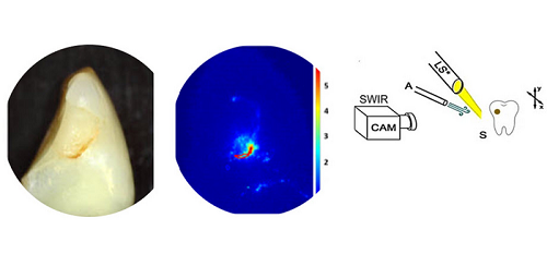
When blended with air drying, scientists employed shortwave-infrared (SWIR) imaging to detect lively dental cavities. This is feasible because active cavities are porous and keep far more drinking water, which affects the infrared measurements all-around the impacted area as the tooth dries. Courtesy of Chang et al., doi: 10.1117/1.JBO.28.9.094801.
The researchers examined 63 extracted human enamel with a complete of 109 suspected secondary lesions. They measured the thickness of the clear floor layer at the lesion interfaces, which is indicative of lesion activity, employing OCT. They further more verified lesion severity and composition applying MicroCT. OCT and MicroCT measurements of lesion construction, depth, and severity were correlated with fluid decline fees calculated with the SWIR reflectance and the thermal imaging approaches to figure out whether SWIR and thermal imaging ended up indeed valuable for detecting energetic lesions.
Total, SWIR executed better than thermal imaging in the assessment of secondary caries lesions on tooth coronal surfaces, even though both methods done greatest on sleek area lesions. SWIR imaging at 1950 nm was beneficial for differentiating composite restorations and lesions from seem tooth composition. Irrespective of the advanced geometry and topography in a couple of lesions, SWIR reflectance imaging for the duration of dehydration was capable to evaluate the permeability of the tooth with somewhat close correlation to OCT.
Thermal imaging carried out nicely in pinpointing crevices amongst composite substance and tooth structure, but at periods it was masked by the advanced topography of the tooth. SWIR imaging did not surface as vulnerable to this sort of interference, owing to the ability of SWIR to differentiate composite resources, tooth constructions, and lesions with significant contrast.
The SWIR permeability measurements were very well correlated with OCT measurements of the thickness of the clear area layer of the lesions. Escalating clear floor layer thickness led to lowered permeability of lesions and possibly indicated full lesion arrest when it arrived at a thickness equal to or higher than 70 μm.
In accordance to the researchers, nondestructive SWIR reflectance and OCT imaging could give, for the to start with time, a way to detect secondary cavities when they kind. The conclusions of this study could direct to new methods to diagnostic imaging in dentistry.
“The standard methods relying on tactile sensation through a dental explorer and visible inspection based mostly on texture and color are very subjective and unreliable,” Chang claimed. “However, there is at this time no recognized dental imaging technological know-how that can provide diagnostic info with substantial specificity and sensitivity when assessing dental decay exercise.”
Over and above enhanced diagnostics, the perform supports the enhancement of effortlessly operable medical equipment, Chang claimed.
The study was published in Journal of Biomedical Optics (www.doi.org/10.1117/1.JBO.28.9.094801).